Sight - The Eye II Crossword Puzzle
Download and print this Sight - The Eye II crossword puzzle.
Related puzzles:
QUESTIONS LIST:
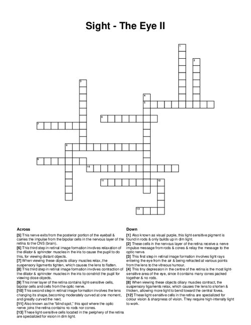
- optic disc : also known as the “blind spot,” this spot where the optic nerve joins the retina contains no rods nor cones.
- rhodopsin : also known as visual purple, this light-sensitive pigment is found in rods & only builds up in dim light.
- accommodation : this second step in retinal image formation involves the lens changing its shape, becoming moderately curved at one moment, and greatly curved the next.
- optic nerve : this nerve exits from the posterior portion of the eyeball & carries the impulse from the bipolar cells in the nervous layer of the retina to the cns (brain).
- refraction : this first step in retinal image formation involves light rays entering the eye from the air & being refracted at various points from the lens to the vitreous humour.
- cones : these light-sensitive cells in the retina are specialized for colour vision & sharpness of vision. they require high-intensity light to work.
- nervous : this inner layer of the retina contains light-sensitive cells, bipolar cells and cells from the optic nerve.
- rods : these light-sensitive cells located in the periphery of the retina are specialized for vision in dim light.
- distant : when viewing these objects ciliary muscles relax, the suspensory ligaments tighten, which causes the lens to flatten.
- constriction : this third step in retinal image formation involves contraction of the dilator & sphincter muscles in the iris to constrict the pupil for viewing close objects.
- close-up : when viewing these objects ciliary muscles contract, the suspensory ligaments relax, which causes the lens to shorten & thicken, allowing more light to bend toward the central fovea.
- fovea centralis : this tiny depression in the centre of the retina is the most light-sensitive area of the eye, since it contains many cones packed together & no rods.
- enlarges : this third step in retinal image formation involves relaxation of the dilator & sphincter muscles in the iris to cause the pupil to do this, for viewing distant objects.
- bipolar : these cells in the nervous layer of the retina receive a nerve impulse message from rods & cones & relay the message to the optic nerve.
